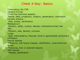
Copy and paste the code below into your blog post or website
Copy URL
Embed into WordPress (learn more)
Comments
comments powered by DisqusPresentation Slides & Transcript
Presentation Slides & Transcript
Chest X-Ray: Basics
Interpreting the CXR
General format:
Check the technical aspects
-Name, date, projection, rotation, penetration, inspiration
Cardiac shadow
-Size, shape, calcification
Mediastinum
-Position, width, outline, density, tracheobronchial tree
Hila
-Position, size, density, concave
Lungs
-Size, transradiancy, fissures, focal or generalised pulmonary infiltration
Pleural spaces
-Effusions, soft tissue masses, calcification, pneumothorax
Bones
-Fractures, lytic or sclerotic lesions
Soft tissues
-Masses, calcification
Technical Aspects
Check the side markers
Have you got L and R the right way around?
Assessment of rotation
Distance between medial ends of the clavicles and the spinous processes
Penetration
• Intervertebral disc spaces just visible through mid-cardiac shadow
• Pulmonary structures visible with clarity
• Few vessels in outer third
Inspiration
Full inspiration:
• 6-7 anterior ribs
• 10-11 posterior ribs
Cardiac shadow
Heart Size
• Cardiac Transverse Diameter (CTD) = a+b
< 15.5cm (males)
< 15.0cm (females)
• Cardio-Thoracic Ratio (CTR) =a+b÷c+d
< 0.5
Cardiac configuration
• Dilated left ventricle:
-Ischaemic/dilated cardiomyopathy
-Aortic reflux
-Mitral reflux
-VSD, PDA
-Anaemia, hyperthyroid, Paget’s, AVF
• Failing left ventricle:
-AS, HTN, Coarctation
• Cardiomegaly (CTR > 0.5)
• Large third “mogul”
• Double density right heart border
• Displaced descending aorta
• Dilated left atrium:
-Mitral stenosis or myxoma
-Mitral reflux, VSD, PDA,
-ASD (late)
The mediastinum
• Mediastinal position
• Ratio:
- 1/3 to the Rt of midline
- 2/3 to the Lt of midline
Left heart border
• Aortic arch
•Main (left) PA
•Left atrial appendage
•Left ventricle
Right heart border
•Brachiocephalic vein
•SVC
•Right atrium
•IVC
- blurred or obscured and 'indistinct' right heart border
Hila
•Position of hilar point:
-Right = 6th rib in mid-axillary line
-Left = 0–2.5cm higher
•Outline:
- "V" shaped
Lungs
•Size
•Transradiancy
•Fissures
•Focal or generalised pulmonary infiltration
Horizontal fissure
6th rib in mid-axillary line
Azygos fissure
4-layers of pleura, containing azygos vein
Plural Spaces
•Effusions
•Soft tissue masses
•Calcification
•Pneumothorax
Bones
•Fractures
•Lytic or sclerotic lesions
Soft Tissues
•Masses
•Calcification